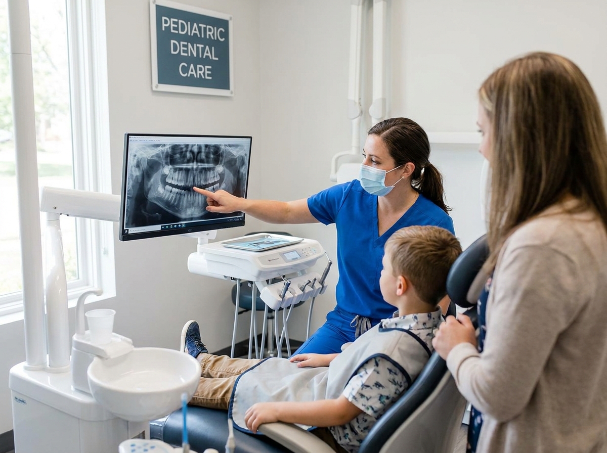
How to Ensure Safe Dental X-Rays for Children: Complete Safety Guide
Dental X-rays for children involve minimal radiation exposure when performed according to pediatric radiology guidelines. According to the American Academy of Pediatric Dentistry (2024), digital dental imaging exposes children to approximately 80% less radiation than traditional film X-rays, making modern pediatric dental radiography safer than ever before.
Understanding Pediatric Dental X-Ray Safety and Radiation Exposure
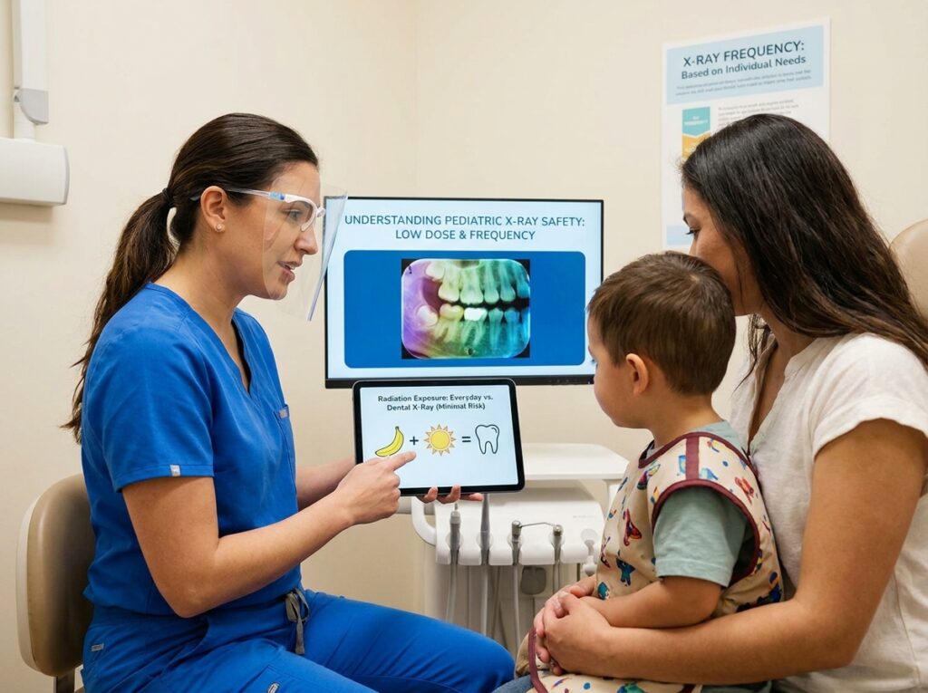
The Food and Drug Administration (FDA) confirms that children’s dental X-rays use ionizing radiation measured in microsieverts, with a typical bitewing series delivering 5 microsieverts—equivalent to one day of natural background radiation. A pediatric dentist applies the ALARA principle (As Low As Reasonably Achievable) to minimize radiation exposure children receive during diagnostic imaging.
Digital radiography has revolutionized safe X-rays for kids by reducing radiation dose by 50-80% compared to film X-rays. Modern X-ray machines in pediatric dental clinics use focused beams and protective equipment to ensure radiation safety during every oral examination technique.
Before You Begin: Prerequisites for Safe Pediatric Dental Imaging
- Verify clinical necessity: Confirm with your pediatric dentist that X-ray benefits outweigh minimal radiation exposure for your child’s specific oral health needs
- Choose digital imaging: Ensure the dental office uses digital X-rays vs film for low radiation X-rays
- Review medical history: Inform the dental hygienist about previous radiographic examinations within the past 6 months
- Confirm protective measures: Check that lead apron and thyroid collar are available in appropriate pediatric sizes
- Prepare your child: For children with dental anxiety, review our guide on how to help a child overcome dental anxiety
Types of Dental X-Rays Used in Pediatric Dentistry
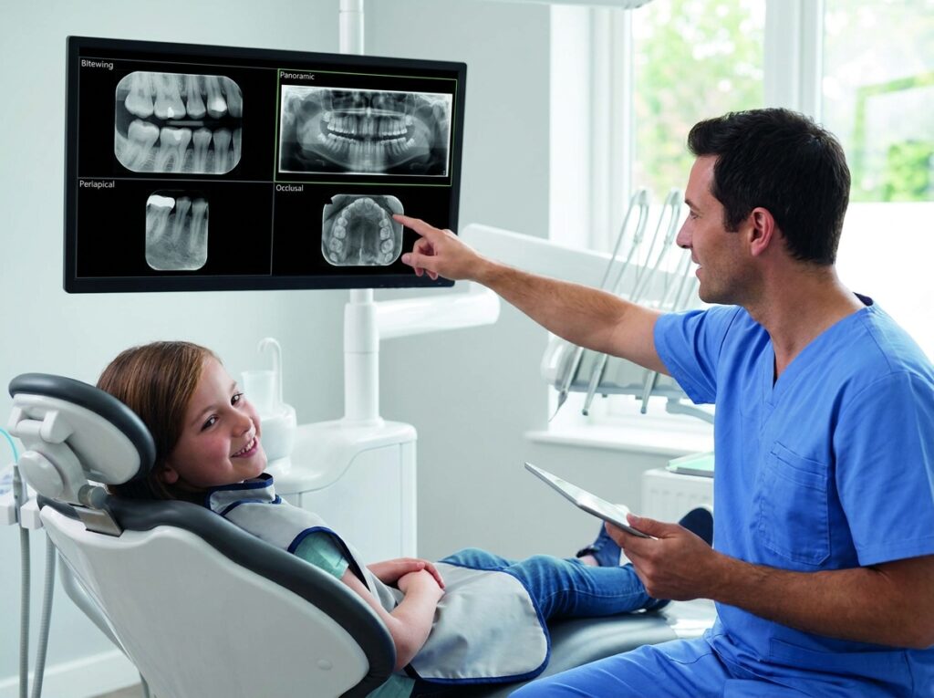
A radiologist or pediatric dentist selects from several diagnostic dental imaging types based on tooth development stages and diagnostic needs:
| X-Ray Type | Purpose | Typical Age |
|---|---|---|
| Bitewing X-rays | Cavity detection between teeth, monitors enamel and dentin health | Age 4+ |
| Periapical X-rays | Shows entire tooth structure from crown to root, detects dental abscess and bone loss | As needed |
| Panoramic X-rays | Captures jaw development, tooth eruption patterns, impacted teeth, skeletal development | Age 6+ |
| Cone beam CT (CBCT) | 3D imaging for complex cases, orthodontic treatment planning, identifies malocclusion | Specialized cases only |
Step-by-Step Safety Protocol for Children’s Dental X-Rays
Step 1: Confirm Medical Necessity and Timing (5 minutes)
The dental office staff reviews your child’s oral health screening history. The American Dental Association (ADA) recommends X-ray timing based on individual risk factors rather than routine dental X-rays at fixed intervals. High-risk children with active tooth decay may need bitewing radiographs every 6 months, while low-risk children may wait 12-24 months between imaging sessions.
Step 2: Position Protective Equipment (2 minutes)
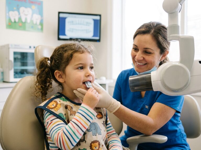
The radiation safety officer or trained staff member places a lead apron children can wear comfortably across the chest and abdomen. A thyroid shield wraps around the neck to protect the thyroid gland. According to FDA guidelines (2023), these radiation protection measures reduce scattered radiation exposure by 95%.
Step 3: Optimize Machine Settings (1 minute)
The dental hygienist adjusts the X-ray machine to pediatric-specific settings, reducing millisieverts to age-appropriate imaging levels. Modern dental technology automatically calibrates low-dose imaging parameters for infant dental X-rays, toddler X-rays, preschool dental imaging, school-age X-rays, and teen X-rays.
Step 4: Conduct Rapid Image Capture (30 seconds per image)
Digital dental imaging requires exposure times of 0.1-0.2 seconds per radiograph—significantly faster than historical film X-rays that required 0.5-1.0 seconds. Quick imaging protocols minimize movement artifacts in young children.
Step 5: Review Images Immediately (2 minutes)
The pediatric dentist performs radiographic interpretation on-site using dental diagnostic tools. Immediate review confirms adequate diagnostic benefits without requiring repeat exposures that increase cumulative radiation dose.
Recommended Dental X-Ray Schedule for Children by Age and Risk Level
The American Academy of Pediatric Dentistry (AAPD) publishes imaging protocols based on cavity risk assessment. The recommended X-ray schedule varies by developmental stage:
Primary Teeth Stage (Ages 2-6)
Children with baby teeth typically receive their first dental X-ray when posterior teeth contact each other (usually age 4). Low-risk patients may wait until age 5-6, while high-risk children with visible dental caries need baseline X-rays at the initial comprehensive examination.
Mixed Dentition Stage (Ages 6-12)
During the transition from primary teeth to permanent teeth, bitewing X-rays monitor tooth development and detect cavities. Children without dental problem diagnosis may need bitewings every 12-24 months. Those with active decay require 6-12 month recall X-rays for childhood dental care monitoring.
Adolescent Stage (Ages 13-18)
Teens typically need biannual X-rays if cavity-prone, or annual dental X-rays if low-risk. Adolescent dental X-rays before orthodontic treatment include panoramic dental X-rays to assess jaw X-rays and wisdom tooth development.
Radiation Risk Assessment: Understanding Safe Exposure Limits
Parents often ask are dental x-rays safe for kids due to concerns about cancer risk. According to the FDA (2024), the radiation exposure from a full-mouth series of pediatric radiographs (approximately 35 microsieverts) equals 3-4 days of natural background radiation or one cross-country flight. The lifetime cancer risk from a single pediatric dental X-ray series is estimated at less than 1 in 2 million—far lower than the risk of untreated tooth decay.
Troubleshooting Common Pediatric X-Ray Challenges
If Your Child Experiences Anxiety During Imaging
Solution: Request a practice session where your child sits in the chair without actual X-ray exposure. Use distraction techniques or allow parents to remain visible during imaging. Some pediatric dental clinics offer sedation dentistry for extremely anxious children requiring emergency dental X-rays.
If Images Appear Blurred from Movement
Solution: Digital radiography allows immediate retakes with minimal additional exposure. Orthodontists and oral surgeons often use bite blocks or positioning aids to stabilize young children’s heads during tooth imaging.
If Your Child Gags During Sensor Placement
Solution: Smaller pediatric sensor sizes reduce gagging. Breathing through the nose and salt on the tongue can minimize gag reflex during non-invasive diagnostics.
If You Want to Refuse or Delay X-Rays
Solution: Discuss imaging alternatives and clinical necessity with your pediatric dentist. While parents can decline radiographs, the dentist cannot diagnose interproximal cavities or root canal problems without diagnostic imaging children can receive safely. Most dental insurance requires current radiographs for treatment authorization.
Next Steps for Ongoing Dental Health Monitoring
After establishing your child’s baseline radiographs, schedule follow-up imaging according to their individualized dental X-ray frequency plan. Maintain preventive dental care through:
- Regular checkup X-rays as recommended by your pediatric dentist (typically 6-24 months)
- Fluoride treatments to strengthen enamel and reduce future X-ray needs through tooth decay prevention
- Dietary counseling to minimize cavity development requiring frequent diagnostic dental imaging
- Sealant application on permanent molars to reduce early cavity detection needs
- Orthodontic evaluation by age 7 using panoramic X-rays if growth patterns suggest malocclusion
Modern pediatric dental radiography equipment and radiation minimization safety precautions ensure children receive optimal diagnostic benefits with negligible health risks. By following age-appropriate X-ray protocols and safety guidelines, parents can confidently support their children’s oral health screening needs throughout development stages.